
Featured
Leveraging Video & GenAI for Enhanced Visual Workflow Efficiency
By optimizing AI workflow automation with AI, video and AWS-powered solutions, Cardinal Peak helped WorkDone cut processing costs by 99%, enhance procurement efficiency and scale seamlessly. Read the full case study to see how artificial intelligence-driven automation transforms business processes.
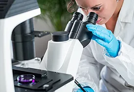
Transforming Scientific Discovery with AWS Cloud-Based Digital Image Processing
Faced with customers filling terabyte drives and struggling to collaborate, our client needed to centralize storage, accelerate processing and enable seamless teamwork. Discover how Cardinal Peak built a multitenant cloud solution on AWS that streamlines digital image processing and facilitates easy collaboration through cloud access.

Remote Surveillance Camera System Development to Modernize Investigative Surveillance
In this case study, Cardinal Peak’s video product design and development expertise unlocked remote accessibility and significantly more efficient intel surveillance, helping one of New Jersey’s largest private investigation firms realize an astounding 80% profit boost.
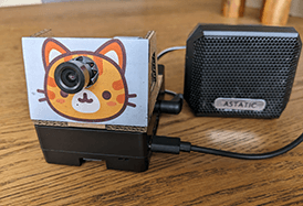
Developing an Amazon Kinesis-Powered Cat Detector: An Edge Machine Learning Case Study
This case study reveals the secret to outsmarting mischievous feline antics with our innovative cat detector powered by Amazon Kinesis Video Streams, AWS IoT Greengrass and machine learning.
Faced with customers filling terabyte drives and struggling to collaborate, our client needed to centralize storage, accelerate processing and enable seamless teamwork. Discover how Cardinal Peak built a multitenant cloud solution on AWS that streamlines digital image processing and facilitates easy collaboration through cloud access.
In this case study, Cardinal Peak’s video product design and development expertise unlocked remote accessibility and significantly more efficient intel surveillance, helping one of New Jersey’s largest private investigation firms realize an astounding 80% profit boost.
This case study reveals the secret to outsmarting mischievous feline antics with our innovative cat detector powered by Amazon Kinesis Video Streams, AWS IoT Greengrass and machine learning.