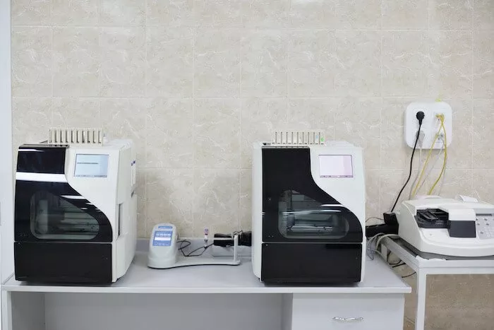
 Medical Imaging is familiar to most people and recognized as a critical non-intrusive diagnostic tool. Whether the imaging takes the form of X-rays, ultrasound images, MRI scans or CT scans, most people have had an imaging procedure performed and understand the diagnostic value they provide to the doctor or surgeon. These same benefits are important to the nurses, doctors, and surgeons who have animals as their patients. Your dog may have an obstruction in his stomach, or your prize racehorse is having problems during pregnancy, or the California condor at your local zoo must be saved from extinction – for these creatures imaging is just as critical as it is for humans.
Medical Imaging Device Development for Field Veterinarians
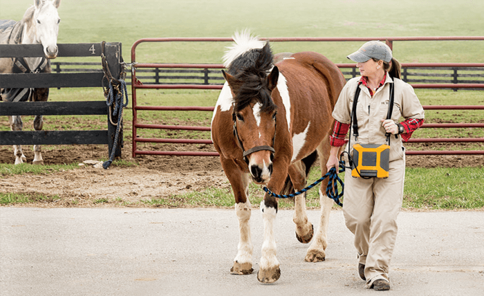
While there are many possible animal patients for medical imaging, the drivers in the industry include livestock producers, racehorse owners and breeders, and large zoos and preserves. The animals in these businesses carry a lot of value. Their health, well-being, and breeding ability is key to the business profitability and in some cases the future of the animal species itself. The businesses operate in a time-sensitive environment where a missed breeding cycle or worse, loss of life, is unacceptable.
Cardinal Peak has been an extension of our engineering capabilities for three generations of our products. They are knowledgeable and great to work with. They have been a long-term solution for us.
 The working environment for a field veterinarian is very different than that of a doctor in a clinic handling human aches and pains and minor outpatient surgery. Livestock production facilities are large, dirty, cramped places with no resemblance to a sterile doctor’s office. The working surface is usually the ground itself, covered with hay and other undesirable material. There may be few electrical power sources, poor visibility, and extreme temperature conditions. To work in this environment, any medical imaging device must be portable, rugged, reliable, accurate, lightweight, and cost-effective.
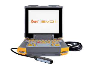
Designing a Portable Ultrasound Video Solution
E.I. Medical Imaging came to Cardinal Peak looking for a strong software team with extensive experience in video processing. They brought with them a custom platform, looking much like a ruggedized laptop, as the basis for a new portable ultrasound imaging device specifically designed for veterinarians. The ultrasound images are captured by a variety of probes and viewed in real time on the screen. As the probe is repositioned on the animal, the video must instantly update to provide relevant information. Low latency and high resolution are critical attributes of the ultrasound imaging device. E.I. Medical found the team they were looking for at Cardinal Peak.
The Cardinal Peak software team was responsible for all the embedded healthcare software including the video processing of the ultrasound image. We started with a set of requirements from E.I. Medical and architected the software solution. We wrote the code and performed unit testing and functional testing. The embedded software controls all aspects of the ultrasound device and accommodates the many different types of probes used by the veterinarians. A new user interface was designed to make the device as easy to use as possible, to speed the scanning process, and reduce the stress on the animal being examined.
Ultrasound Medical Device Development Challenges
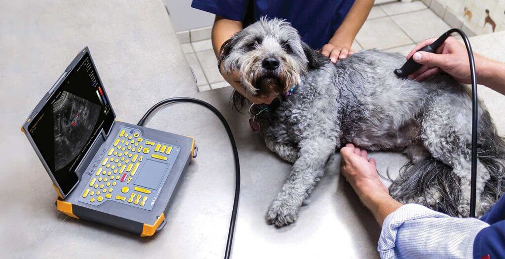 Processing the video image represented the biggest technical challenge in the software design. The video is continuously captured by the ultrasound probe and must be displayed on the screen with very low latency. The image must update instantaneously as the probe is repositioned during the scanning process. Critical diagnostic information would be lost if the display was slow to update or the image was distorted, blurry, or contaminated with artifacts. Details are important, so the image needed to be high-resolution with 60 frames per seconds.
To overcome these challenges the software team immersed themselves in the details of the hardware design provided by E.I. Medical. Careful attention was paid to the CPU interface to memory, and the read/write speeds of the various types of memory available to the CPU. The software was carefully architected to take advantage of local RAM whenever possible to significantly reduce the time lost during data transfer. Algorithms were carefully crafted to match the hardware design and optimized to run as fast as possible. The cross-discipline engineering skills of the Cardinal Peak software developers was key to meeting the demanding requirements of ultrasound image processing on a small, portable, long-battery-life product.
Some interesting features were included in the software development:
- Multiple imaging modes are supported
- Record and playback of an 8 second buffer
- Ability to stream the video to a secondary monitor or phone
- Viewing the video on a video headset in high-light environments
The ultrasound device from E.I. Medical is a success in the marketplace because it allows the veterinarian to provide a high quality of care to animals large and small. Cardinal Peak is proud to have contributed to the software development and helped E.I. Medical become the leader in portable veterinarian ultrasound products.
Check out our informative blog post showcasing diverse machine learning for signal processing applications. Dive in to navigate this exciting intersection of technologies.